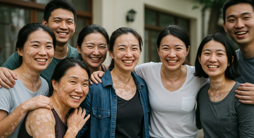
White or pale patches on the skin do not always mean you have vitiligo. Several other skin conditions can look similar. Getting the right diagnosis matters because each condition has a different cause and requires a different treatment approach.
Misdiagnosis of vitiligo is not uncommon, especially in the early stages when patches are small, or on lighter skin tones where the contrast is less obvious. If you have noticed unexplained changes in your skin colour, a dermatologist is the right person to make an accurate diagnosis.
Vitiligo and albinism are commonly confused because both can result in very pale or white skin. However, they are fundamentally different in their cause, appearance, and management:
The most obvious visual difference between vitiligo and albinism is distribution. Vitiligo causes depigmentation in patches. Some areas of skin retain their natural colour while others lose it entirely and turn white.
On the other hand, albinism causes full lightening across the whole body from birth. A person with albinism will have consistently pale skin, hair, and eye colour throughout their life.
Vitiligo is classified as an autoimmune condition. The individual’s immune system mistakenly targets and destroys functioning melanocytes in specific areas of the skin.
Albinism, by contrast, is a genetic condition present from birth. The body’s melanocytes exist in normal numbers but carry mutations that prevent them from producing melanin correctly. This is why the two conditions require very different treatment approaches.
In Singapore, where many people have medium to deeper skin tones, vitiligo patches are often highly visible against surrounding pigmented skin. Albinism is immediately apparent from birth and is unlikely to be confused with vitiligo in a clinical setting.

| Vitiligo | Albinism |
|---|---|
|
|
Tinea versicolor, also known as pityriasis versicolor, is another skin condition often confused with vitiligo. Both can produce lighter patches on the skin, and both are common in tropical climates like Singapore’s.
The root causes for each condition are entirely different. Tinea versicolor is attributed to the overgrowth of Malassezia, a type of yeast that lives naturally on almost everyone’s skin.
In some people, particularly in hot and humid conditions like Singapore, the yeast multiplies and interferes with normal skin pigmentation, producing patches that can be lighter or darker than the surrounding skin. It is a surface-level infection.
Vitiligo, meanwhile, involves the immune system destroying the cells responsible for pigmentation.
Tinea versicolor responds best to antifungal treatment, such as topical antifungal creams, antifungal shampoos, or oral antifungal medication. The infection will clear, though it may recur, and skin colour will take some time to return fully.
Vitiligo does not respond to antifungal treatment, and treating it for tinea versicolor will have no effect. This is why professional diagnosis is essential before beginning any treatment.
| Vitiligo | Tinea Versicolor |
|---|---|
|
|
Leukoderma is not a separate condition, but a broad term referring to white skin or loss of skin pigment. In other words, vitiligo is a form of leukoderma. When people use the word leukoderma, they usually mean chemical leukoderma specifically.
Chemical leukoderma is depigmentation caused by physical exposure to specific chemicals, mainly industrial compounds found in rubber, plastic, and certain adhesives and sealants.
These chemicals damage or destroy melanocytes in the affected area, creating white patches that may appear similar to vitiligo.
On the other hand, vitiligo is an autoimmune disease in which the body’s own immune system mistakenly attacks healthy melanocytes, causing pigment loss in the skin.
| Vitiligo | Chemical Leukoderma |
|---|---|
|
|
Appearance alone is not enough to distinguish vitiligo from other conditions that cause white or depigmented skin. To determine what condition you may have, consider the following:
A family history of vitiligo increases your risk of developing it. Albinism is directly inherited, but tinea versicolor has no hereditary element.
Vitiligo can be triggered by stress and hormonal changes in people who are already genetically susceptible to the condition.
Emotional stress is one of the most commonly reported triggers. Periods of anxiety, grief, or major life changes can precede the onset or flare-up of the condition. Physical stress on the skin, such as sunburn, injuries, and chemical exposure, can also trigger new patches.
Hormonal changes during puberty or pregnancy may also play a role in triggering the condition in some individuals. It is important to note that triggers do not cause vitiligo on their own, but only activate it in people who already have an underlying predisposition.
Vision problems or extremely pale eye colour from birth strongly suggest albinism rather than vitiligo.
Lighter patches may appear on your skin after healed eczema, psoriasis, or a wound. These could be signs of post-inflammatory hypopigmentation rather than vitiligo.

See a dermatologist promptly if you notice any of the following:
If your diagnosis is vitiligo, starting treatment early gives the best chance of slowing progression and achieving repigmentation.
If it is tinea versicolor or another condition, appropriate treatment can resolve it more quickly and prevent complications. Do seek professional assessment as soon as possible for any noticeable changes in your skin.
Different conditions require different diagnostic approaches. A dermatologist will use a combination of clinical observation, tools, and tests to reach an accurate diagnosis. Here is what you can expect.
A Wood’s lamp, also known as a blacklight test, is a handheld ultraviolet light used in a darkened room to examine the skin. It is one of the most useful tools for distinguishing vitiligo from other skin conditions, and it is painless and non-invasive.
Under a Wood’s lamp, different conditions will produce different results:
| Condition | Appearance Under Wood’s Lamp |
|---|---|
| Vitiligo | Bright, chalk-white colour with clear borders around the patches |
| Tinea versicolor | Fluorescent yellow-orange colour |
| Albinism | Uniform, faint-white skin, blurred edges (in partial albinism) |
| Other bacterial or fungal infections | Typically brightly-coloured (blue, green, pink) |
While it works for all skin tones, the Wood’s lamp is particularly useful on lighter skin, where vitiligo patches may be difficult to see in natural light. The bright fluorescence makes skin patch boundaries clearly visible, which also helps doctors evaluate the extent of the condition.
Your dermatologist will examine the size, shape, distribution, and borders of patches. They will assess whether the depigmentation is complete or partial, the placement of depigmented areas, and the overall condition of your skin.
Your doctor will also take a detailed personal history: when the patches appeared, whether they are spreading, your family history, occupation, skincare products, and any associated conditions, to determine what skin condition you may have.
A dermoscope is a small handheld device that allows a doctor to examine the skin up close. In vitiligo patches, it can reveal tiny dots of residual colour around the hair follicles. These dots indicate that some melanocytes are still alive, which means the skin may respond well to treatment. This pattern is rarely seen in other conditions that cause pale or white patches, making it valuable in confirming a vitiligo diagnosis.
In some cases, additional tests are needed to confirm a diagnosis. Your doctor may want to examine your skin cells under a microscope to observe the presence of melanocytes or bacteria. They may also order a blood test to check for related autoimmune conditions, like thyroid diseases, which are more common in people with vitiligo.
A skin biopsy may be taken if the diagnosis remains uncertain after clinical examination and Wood’s lamp assessment. This can help confirm the absence of melanocytes, which indicates vitiligo, or determine if another skin condition is present.
Getting an accurate diagnosis is the most important step toward getting the right help. Whether your condition turns out to be vitiligo or something else entirely, a correct diagnosis means you can start the right treatment without delay.
Yes, vitiligo can be misdiagnosed, particularly in its early stages or in people with lighter skin tones who show less contrast between affected and unaffected skin.
Conditions most commonly confused with vitiligo include tinea versicolor and chemical leukoderma. In some cases, vitiligo can be mistakenly treated with antifungal medication when tinea versicolor is assumed, or vice versa. Misdiagnosis can lead to delays in appropriate treatment and unnecessary use of ineffective therapies.
A dermatologist with experience in pigmentation disorders is best placed to make an accurate diagnosis. If you have been receiving treatment for a skin condition without improvement, it can be beneficial to seek a second opinion.
Vitiligo is typically confirmed through a combination of clinical examination, Wood’s lamp assessment, and other lab tests if necessary.
A dermatologist will examine the pattern, distribution, and appearance of the patches, and use a Wood’s lamp to check whether the patches fluoresce bright white, which is characteristic of the complete depigmentation seen in vitiligo.
Dermoscopy can be used to reveal surviving melanocytes in the skin patches. If the diagnosis is still uncertain, a skin biopsy can be taken and examined under a microscope. Blood tests may also be ordered to check for associated autoimmune conditions, not to confirm the vitiligo diagnosis itself. The whole process is typically quick, painless, and non-invasive.